Methods of Re-operation

Before surgery all patients undergo a special CT scan of the chest to help us understand the anatomy of the heart and blood vessels and therefore anticipate problems and difficulties during re-operations. The CT scan is able to precisely locate all the cardiac chambers, blood vessels, bypass grafts and heart valves so we can avoid injuring these structures while we open the chest and free-up the heart. He also is able to look at the major arteries to see if they are suitable for "cannulation" for the heart lung machine.
Cannulation of arteries (placing a tube within the artery through which the heart lung machine can pump blood back into the body) is a critical part of a re-operation - not only is this required to conduct the open heart aspect of the surgery, but cannulation of arteries helps mitigate against any problems encountered on entering the chest or freeing up the heart. For some patients, before we open up the chest we will make a small cut underneath one of the collar-bones to perform cannulation to an artery going to the arm. Sometimes we will also cannulate a vein and place the patient on the heart-lung machine before we open the chest making entry into the chest very safe. This way even if injuries to the heart or blood vessels occur when we divide the breastbone they occur in a very controlled situation and the risks are minimal. After the breastbone is divided we free up only the aspects of the heart necessary to expose the heart valve(s) we are operating on. By doing minimal "dissection" in this manner we reduce the risk of injury to the heart and bleeding afterwards. Although most patients have surgery done through an incision in the middle of the chest, sometimes we will perform re-operations through a cut at the side based on findings on the CT scan.
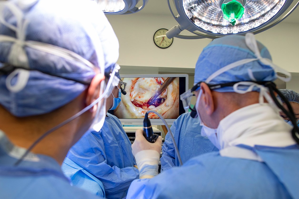
Once we have safely entered the chest and freed up the necessary parts of the heart, we open up the heart to see the heart valve. Dr. Adams, Dr. Anyanwu and team have vast experience in dealing with valve re-operations in various scenarios:
- If the valve has not been operated before then valve repair is undertaken in the same way as for a patient who has not had valve surgery.
- Valve Re-repair: If the valve has been repaired before but is leaking again, Dr Adams and Dr Anyanwu will specifically evaluate the echocardiogram and then examine the valve to determine the reason the previous operation failed. Sometimes we find it is because there has been development of new disease in the valve, sometimes we find there has been a technical failure or flaw from the previous repair. Once the cause of the leak is identified we would re-repair the valve. Usually this will entail us removing any previously implanted rings and then taking down the old repair. We then reconstruct the valve using various techniques depending on what we find. Sometimes we have to use part of the patient's tissue around the heart (pericardium) to recreate a new valve leaflet. We are able to re-repair the majority of valves, especially where the first repair was for valve prolapse. If repair is not possible then we would perform a valve replacement.
- Valve Replacement: If previous replacement has been undertaken, then we will remove the existing artificial valve, and thoroughly clean up the valve annulus - which is the part of the heart where the artificial valve sits and the place a new artificial valve. Thorough cleaning of the valve annulus (removing all previous surgical material, abnormal tissue and scar tissue) is essential to allow the heart tissues to grow into the new valve - without this the valve will be more prone to early failure due to leakages around the valve.