
Commissures
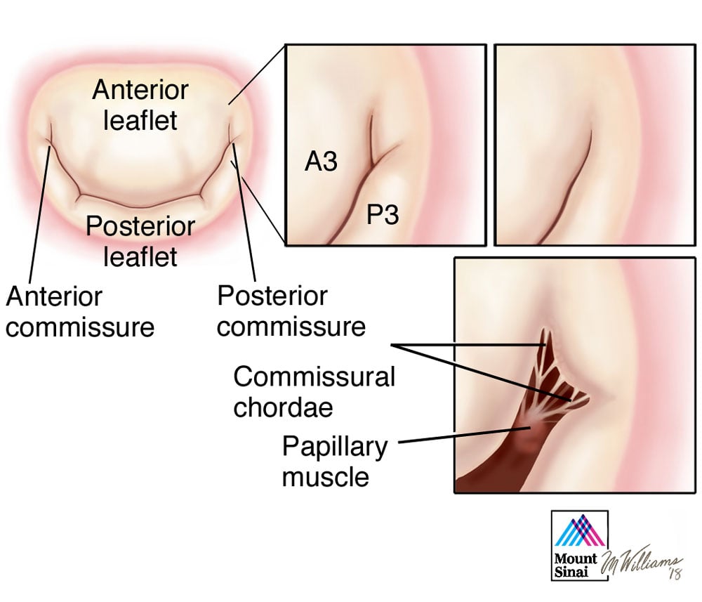
The commissures define a distinct area where the anterior and posterior leaflets come together at their insertion into the annulus. Sometimes the commissures exist as well defined leaflet segments, but more often this area is a subtle structure, and can be identified using two anatomic landmarks: the axis of corresponding papillary muscles and the commissural chordae, which have a specific fan-like configuration. Several millimeters of valvular tissue separates the free edge of the commissures from the annulus. This area must be respected when dealing with prolapse of a commissure and the corresponding anterior and posterior leaflet segment (P3 and A3) by resection, for example, or a residual regurgitation will occur from this region.
* Modified from Carpentier A, Adams DH, Filsoufi F. Carpentier’s Reconstructive Valve Surgery. From Valve Analysis to Valve Reconstruction. 2010 Saunders Elsevier.