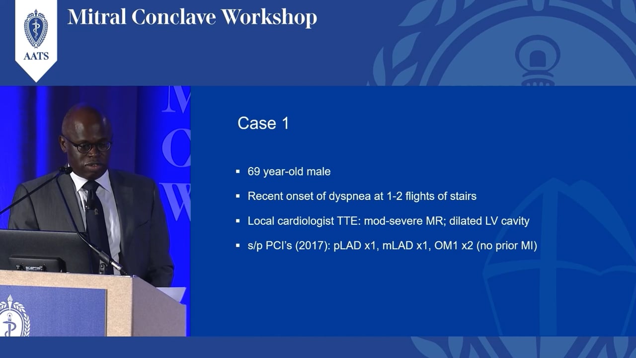
INTRODUCTION:
Good afternoon and welcome back. Hopefully you had a nutritious lunch and we are ready to go again. I am very fortunate to have had the assistance for this entire session from David Adams, Chief from Mount Sinai, who has been very gracious in his participation and assistance; so not only is he going to give us the first talk, which I hope will try and spell out this whole spectrum of degenerative diseases of the valve which we didn’t discuss much last year, and then I am going to allow David to really handle the rest of the session. He has been incredibly helpful at putting together what I think is going to be a very cohesive 2 hours about the mitral valve, so David…
DR. DAVID ADAMS:
Scott, thank you very much. It is my task today to take the next 20 minutes and talk about the spectrum of degenerative diseases affecting the mitral valve by way of disclosure. I am in inventor and a consultant with royalty agreements for Edwards, as well as an unpaid research advisor to Phillips. I would handle that conflict by saying all of the information I am going to cover is available in the peer reviewed literature from our group. Well, I think everybody in this room is familiar with the bookends of degenerative mitral valve disease but I am going to review them quickly with you. This was the original paper from John Barlow describing the syndrome that took on his name, Barlow valve disease, and you can see he is describing this aneurysmal protrusion of the posterior leaflet associated with a mid systolic click and mild mitral regurgitation and it is interesting to note at that point they were making that diagnosis on angiography, not by echocardiography. I think everyone is familiar with the lesions of Barlow valve disease; giant valves with a lot of excess tissue, single segments that can cover the orifice of the valve. It is one of the degenerative diseases that shows restriction of leaflets as well, in this case, an anterior papillary muscle restricting the cleft between P1 and P2 and then microcalcification or fissures that actually progress to true annular calcification. These are all of the lesions that you will see in Barlow valve disease.
The other bookend of differentiating disease is fibroelastic deficiency and this was the original scripture by Professor Carpentier that actually began it. He started to develop this concept in 1975 and presented this paper at the American Heart Association in 1982 where he described basically a different syndrome and said that in addition to the billowing disease of Barlow’s, there was a second entity that had minimal tissue, trans lucid leaflets and very thin chordae associated with chordal rupture and really introduced the concept of fibroelastic deficiency which is something now that we are also all familiar with and here again are the valve and the lesions. You notice in the prolapsing segment you may have ruptured chordae typically and you may have some thickening, but the typical valve has thin chordal rupture, usually single segment and the remainder of the valve is normal and in fact, sometimes really translucent, really thin tissue; so very different than Barlow’s disease.
Carpentier's group went on then to look more carefully at the comparisons between fibroelastic deficiency and Barlow disease and I just took a couple of slides from his paper published with his pathologist, Paul Fornes, in 1999, to show you a couple of the characteristics to keep in mind. The first one when you look at Barlow’s disease, it is a multisegment disease. In fact, half the patient’s they characterize had bileaflet prolapse. In contradistinction, fibroelastic deficiency is basically a disease of the posterior leaflet. The vast majority of patients with fibroelastic deficiency have isolated posterior leaflet prolapse.
The other thing that Carpentier’s group noticed which I think is important in terms of understanding the literature later on, is this age difference. You see, the Barlow patient had an age difference almost 10 years younger than fibroelastic deficiency and the other thing you’ll note in history Barlow’s patients typically have a long history. They find out in their 20’s that they have mitral valve regurgitation and they present for surgery in their 50’s. Fibroelastic deficiency usually presents at an older age with a shorter history and more severe mitral regurgitation.
The other thing you notice is the difference in the valves. The ring sizes were classic rings at the time, and he was modifying some of these to make up for the extra tissue and still, the ring size was averaged 36 in Barlow group, 32 in the fibroelastic deficiency group.
When you look at the valves you need to look at the entire valve. If you look at the prolapsing segment in fibroelastic deficiency, and this is just in 2 recent cases that we have done, histologically it may look the same. You may see myxoid degeneration that you would typically see in a Barlow valve. So you need to look at the entire valve. You see the rest of this valve basically normal and of course, Barlow’s disease is a multisegment disease and they look very differently. They also look very differently on echocardiography and here you can see the typical findings in both: fibroelastic deficiency, thin leaflets, smaller valves, usually a single isolated chordal rupture. Very different than Barlow’s disease: giant annular dilatation, thick billowing leaflets, annular calcification or fibrosis and displacement of the posterior leaflet on to the left atrium. That was characterized many years ago in this paper by Hutchins in 1986 where he noted in Barlows this displacement of the attachment of the mitral valve posterior leaflet away from the atrioventricular group and toward the left atrium. So keep an eye on that when you are trying to distinguish Barlows. It is another useful finding and also something important to address.
My topic was the spectrum of degenerative mitral valve disease. Actually there is not great agreement in the literature and among groups in terms of reading. If you take a look at how people describe disease you will see the confusion. We basically classify valves according to the way Carpentier did, where you classify fibroelastic deficiency as single rupture or thin normal leaflet - usually a single ruptured chord. You can also have fibroelastic deficiency with excess tissue. Typically, it involves the segment that is prolapsing. There can be increased thickness and collagen and you may see myxomatous change. Then you move over to the true myxomatous disease or myxomatous degeneration, and Barlows forme fruste share some characteristics of normal valves and probably the beginning stages of the Barlow deformity. The Barlows valve multisegment are these very large valves with lots of excess tissue. Now, I told you, you could get myxomatous degeneration in this lesion we consider that a secondary lesion and that is why we classify it has FED. Others have classified this as part of the myxomatous spectrum.
I am not sure that matters and it certainly doesn’t for the message I want to give you today. What I want to give you today is the message of excess tissue and how to recognize that as a lesion and how important that is. If you look along this line you can see with FED usually you have minimal leaflet distention. You may have more so, it is a secondary lesion in long standing cases of chordal rupture and then in the form fruste in Barlow’s you begin to see much more excess tissue and we will actually use that to guide our strategy. One of the many important lessons I have learned from working closely with Professor Carpentier is how differentiating the degenerative process can guide your repair strategy. This slide basically summarizes that. I don’t want to get caught up on which technique, I want to get caught up on how to get tissue to guide techniques. So in FED you have less tissue. Preserve the tissue that you have; no resection, minimal resection, limited quadrangular resection, hair cut operation like Chitwood. You pick the technique but limit your resection. Again, in Barlow’s disease, you have excess tissue. You must “remove” tissue and I put remove in quotes. Quadrangular resection, triangular resection, base of leaflet resection, sliding plasty, Fred Morris technique of tying the valve down into the ventricle. Whether you resect or not you must take effective tissue out of the orifice.
Now, I am going to show you a sequence of videos to make this point. I wanted to highlight this one technique that we have used. We call it the Ink Test where after saline testing, as Carpentier described, we mark the closure line with ink. And what that lets you do when you empty the ventricle is look at the coaptation zone or surface below the closure line. It is very useful because, again, service of coaptation is really how we should think about valve repair. So let’s watch these videos now and what I want to do is take you through a sequence of them. Here is a case of fibroelastic deficiency. You can see the power of 3-D echo, single segment prolapse, P3 and again, the sequence is going to take us through no resection to complete resection.
So here is a case for no resection: You have a small valve, minimal tissue in P2. When you have a prolapse in a commisural area with minimal tissue I would really recommend preserving tissue. In this case, we have good secondary chordae so we are going to correct the dysfunction by chordal transfer. So no resection, take several secondary chordae, transfer them to the margin, true size a ring and there is the result. You can see the coaptation surface and how it is preserved by no resection.
Here is a second case: posterior leaflet prolapse, fibroelastic deficiency and in this case you can see that the chordae are thinner. This is not a good case for chordal transfer. The posterior leaflet prolapse, ruptured chord, minimal tissue, a small valve… So in this case instead of chordal transfer we will do Gortex chordoplasty. We will place our Gortex chords in the papillary muscle, pass them through the leaflet, do the final height adjustment after we put a ring in, the ring is small: 26. Minimal tissue, no resection and you see the closure line and how preserving the tissue insures you have a good surface of coaptation.
Here is a third case of fibroelastic deficiency: posterior leaflet prolapsed. You notice in the echo the leaflet is thicker. Here is the case … So there is some distention of the margin of this leaflet. You could treat this with the techniques I have just shown you but we are going to treat this with the limited resection. You have a normal P3 so normal height of the rest of the valve. We have identified 2 good chords. We are going to do a limited triangular resection, a technique very well described at Mayo Clinic and more recently by Irv Kron, an old Carpentier technique. You see, we have done a minimal resection, simple reattachment of the leaflet, size 28 valve and the closure line. So again, respecting tissue - this time some distention - small resection.
Now we are going to move to the fibroelastic deficiency cases that have distended and excess tissue. So here is a case again of posterior leaflet prolapse and you can see I think the difference in this P2 segment that prolapses from the previous 3 cases. So here is a case: a tall P2, 4 cm tall, the adjacent P1 normal, as is P3, a small valve, 32, not a Barlow valve. You see, we are not cutting out the cleft. We are going to preserve part of P2. This is a measured resection just to get the height. We have saved some remnants of P2. A perfect case for annular compression leaflet re-attachment, long surface of coaptation, quadrangular resection.
Here is a second case of FED, tall P2. In this case we are going to do a limited sliding plasty and the reason we are going to do that is to do a fine height adjustment of the residual P2. So again, you don’t take your scissors and cut from here to here – that is going to leave you inadequate tissue. A quadrangular resection should be planned out to preserve tissue. Let me show you. Here is the valve – giant P2 – now, here we have a deep indentation which is usually a good place to favor, so we will pick which side that we are going to preserve of P2 and then go to the indentation. So we will take the indentation out between P2 and P3. Now we are going to lower the height of the residual P2 by a limited resection and there is a triangular based resection as well and you can see as we lower that down a limited sliding plasty will match the residual leaflet height to P3. There it is after a small limited sliding plasty, a small valve again, size 28 and the surface of coaptation.
As we progress along now we are going to show a form fruste case and what you can see here it has some stigmata of Barlow, some excess tissue in the posterior leaflet. It is a fairly short anterior leaflet. Here is the valve. The posterior leaflet has a lot of excess tissue and it is very thick. You see almost the early fissures in the posterior leaflet, but the normal anterior leaflet.. So this is a case for quadrangular resection. We will take out the tallest part of P2. Now we detach the remnants of P1 and P3. It is useful to close this indentation before you do that. Now you see the residual height. It is not a large valve, 32, that why we call that form fruste, good closure line and no risk of SAM with the leaflet height like that.
Here is a second case of form fruste Barlow. In this case you have a prolapse of the anterior leaflet and the severe mitral regurgitation. So you have just chordal elongation, very thick and almost calcified A2, a normal height posterior leaflet. I wouldn’t argue if you said this valve was hard to classify or unclassifiable but we would call it a forme fruste, size 38 ring. We have a lot of excess anterior leaflet tissue compared to the inner commisural distance. We will resect A2 and close the edges and you can see a good line of coaptation so it was a complex valve but a fairly simple repair.
The last cases I will show you are Barlows’ disease. So here is a patient that has the typical stigmata of Barlows’ disease on echo, large valve, excess tissue, thickened leaflets and billowing with displacement of the posterior leaflet. Here is the valve. Large circumference in the posterior annulus, a lot of excess tissue, a larger valve, 38 ring size. So again, we are going to start with quadrangular resection and we will lower the height of what will be the residual posterior leaflet. You can see it is a centimeter, we will reattach that. You do not have to do anything to the anterior leaflet and there is the closure line.
The second Barlow case I will show you: giant valve, a lot of excess tissue and the reason I wanted to show you this case was calcification and restriction. So you see you certainly have a lot of excess tissue and look at this restriction from this cleft from calcification of the anterior papillary muscle. So we resect all of that and cover the residual defect with sliding plasty, a large ring, a good surface of coaptation.
The last case I will show you is the bileaflet Barlow prolapse. You see bileaflet prolapse, excess tissue, ruptured chords in the anterior leaflet, and a very large valve. You can see the fissures in the annulus, a very tall posterior leaflet. The size of the anterior leaflet (that is a 40 sizer) is a very tall valve with a ruptured chord. So we will start by quadrangular resection and we will detach the leaflets. We are also going to do a base resection of the residual leaflet to continue to shorten the height. Then detach the valve to the commissures, reattach it, and check the height. Now we will correct the anterior leaflet prolapse in this case with a triangular resection. It is a very distended margin. Now it really matches the 40 sizer and then we will correct the prolapse with several chordal transfers. Good closure line and a good surface of coaptation.
So in summary, patients today with mitral valve degeneration, I would strongly recommend differentiating the disease, identifying the lesions, respecting tissue, correcting the dysfunctions and preserving the mitral valve. On behalf of my co-authors, I would like to thank you for your attention.
|