
Innovations
Mitral surgeons at The Mount Sinai Hospital are also leading clinical trials that may one day revolutionize less invasive mitral valve repair and replacement.
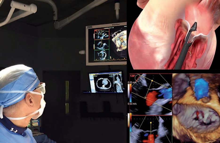
Dr. Adams is the national co-principal investigator of the NeoChord FDA pivotal trial, which is establishing the efficacy of closed beating heart mitral valve repair through a mini-thoracotomy compared with open surgery. In November 2016, Dr. Adams and his team performed the first NeoChord repair procedure in the United States. The NeoChord trial will expand to more than 20 centers in the nation and will be ongoing for the next few years.
In addition, Dr. Adams serves as the national co-principal investigator of the Medtronic Intrepid FDA pivotal trial, which will establish the efficacy of transcatheter closed beating heart mitral valve replacement in patients with increased risk for conventional or surgical mitral valve replacement. This trial began in 2017 and is anticipated to involve more than 40 centers, including The Mount Sinai Hospital, during its course.